cherry angioma images pictures, photos
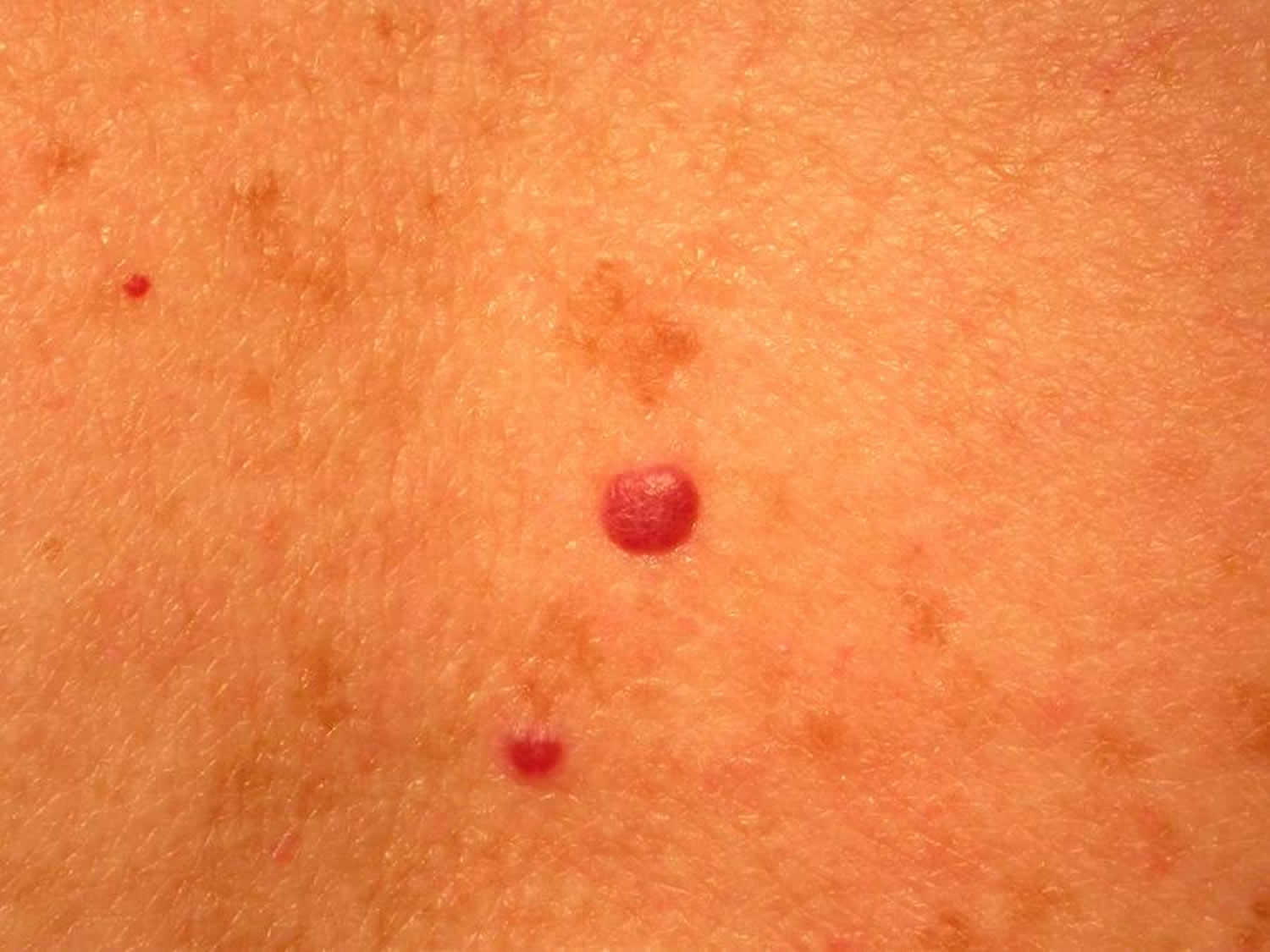
Red moles or cherry angiomas are common non-cancerous skin lesions that can appear as red flat spots or bumps on the skin. They are composed of blood vessels which give them a bright red color hence, giving them the names "red moles" and "ruby spots". Cherry angiomas are often seen in adults over the age of 30 years of age and the elderly.

What are Cherry Angiomas? Dr. Anthony J. Perri
12 Cherry Angioma Pictures. It is a benign (noncancerous) growth on the skin surface that comprises of blood vessels. Many people suffer from these growths at a later stage of their life, with the onset generally occurring at an age of over 40. However, younger people can also get these growths. Picture 1 - Cherry Angioma.
Cherry angioma Symptoms, causes, and treatment
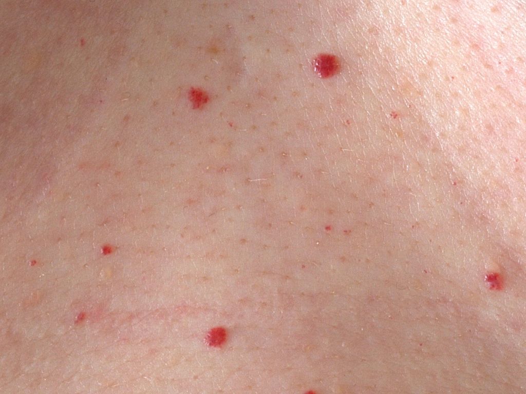
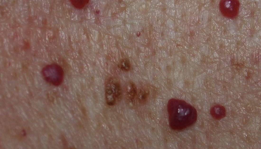
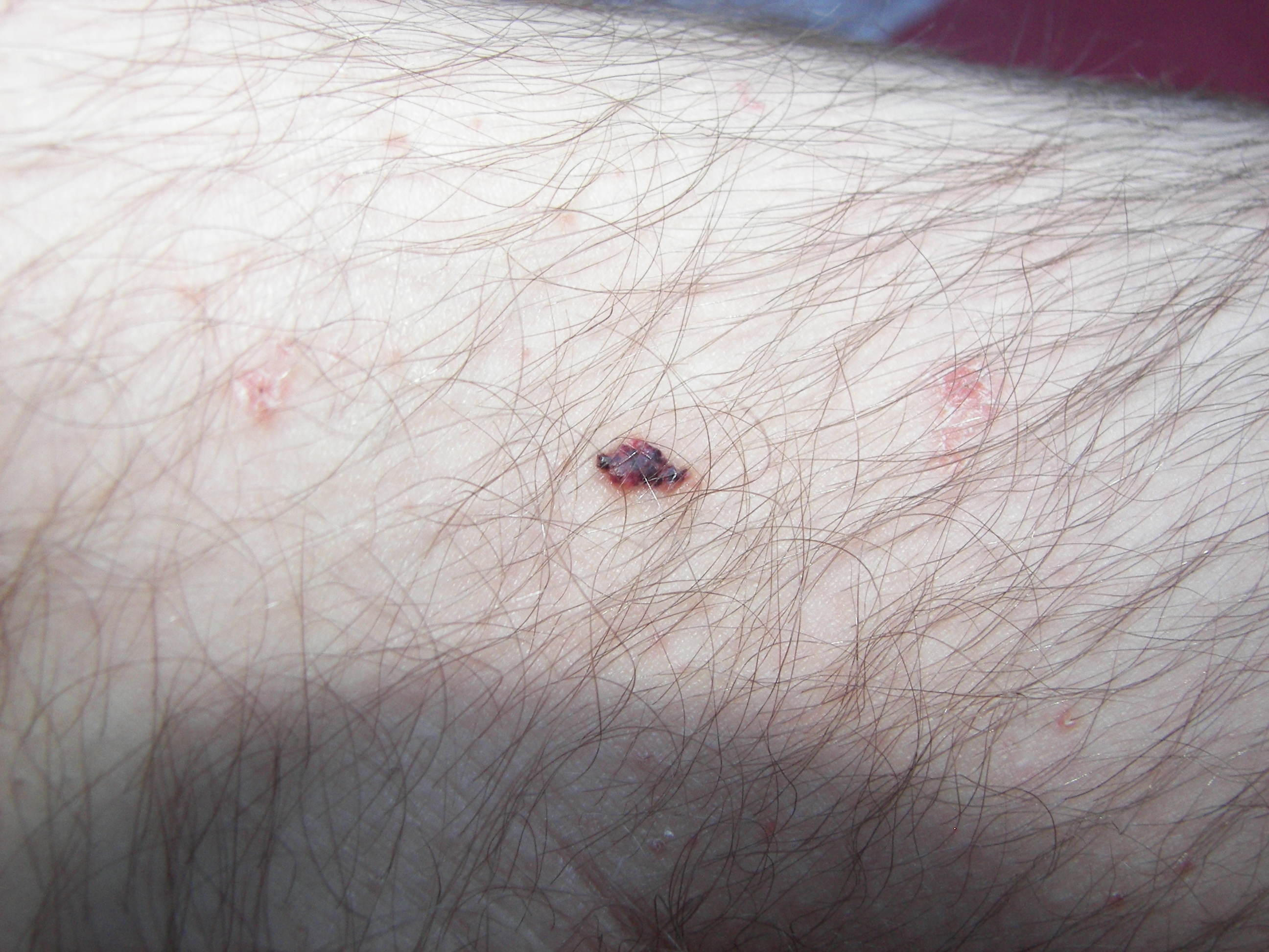
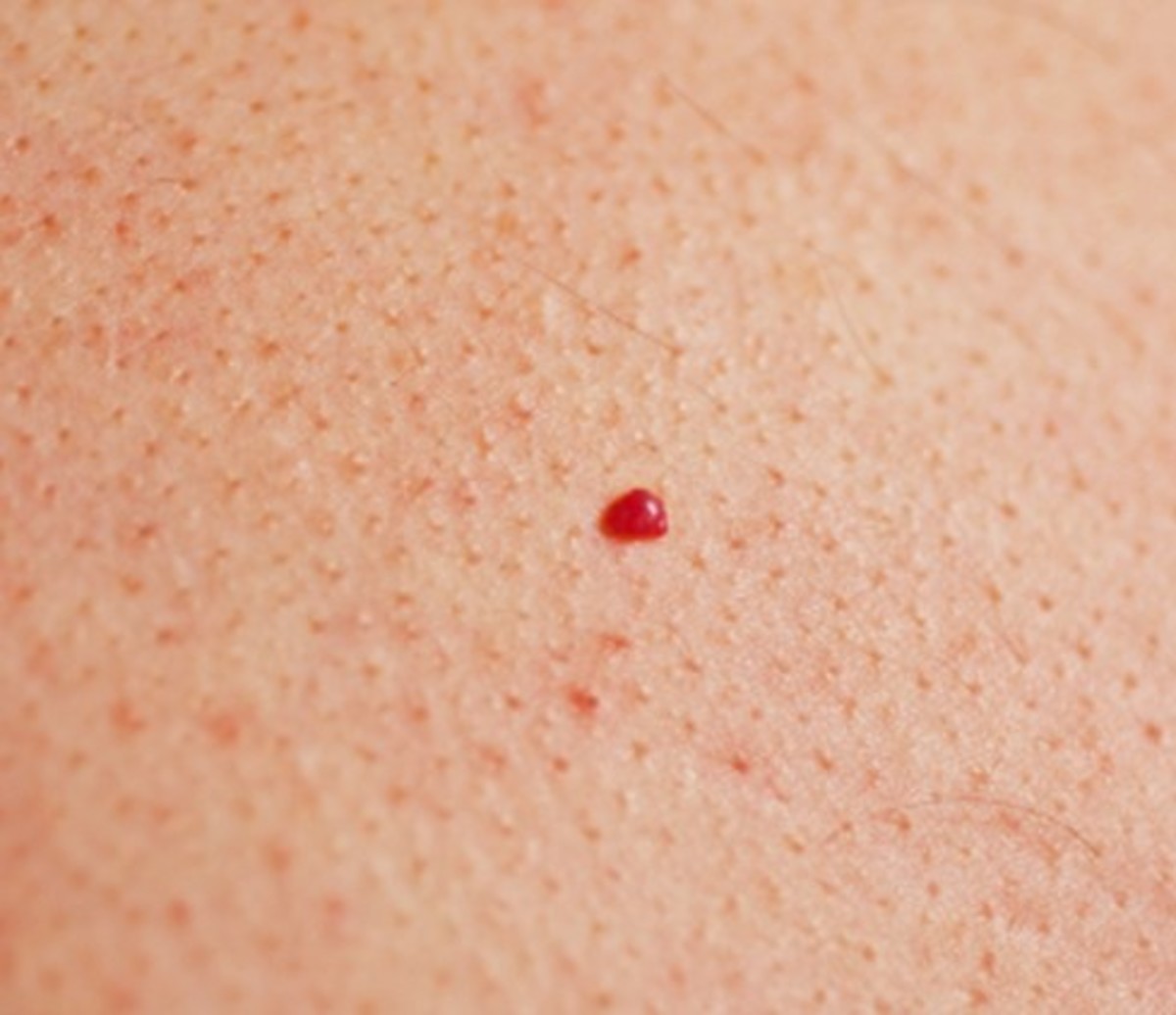
In these cherry angioma pictures, you can see how the angiomas vary slightly in shape and size. Notice how they are all bright red in color, however. Cherry angiomas are most often found on the trunk of the body but they can appear anywhere, including on the arms, legs, neck, and face. The condition is most common in adults over the age of 30.
Cherry Angiomas Pictures, Symptoms, Causes, Treatment, Removal HealDove
If the angioma is large, the doctor may shave off the spot and electrocauterize the skin beneath. Alternatively, they may recommend cryosurgery or CO 2 laser surgery. Cryosurgery refers to when a.
Cherry Angiomas Pictures, Symptoms, Causes, Treatment, Removal HubPages
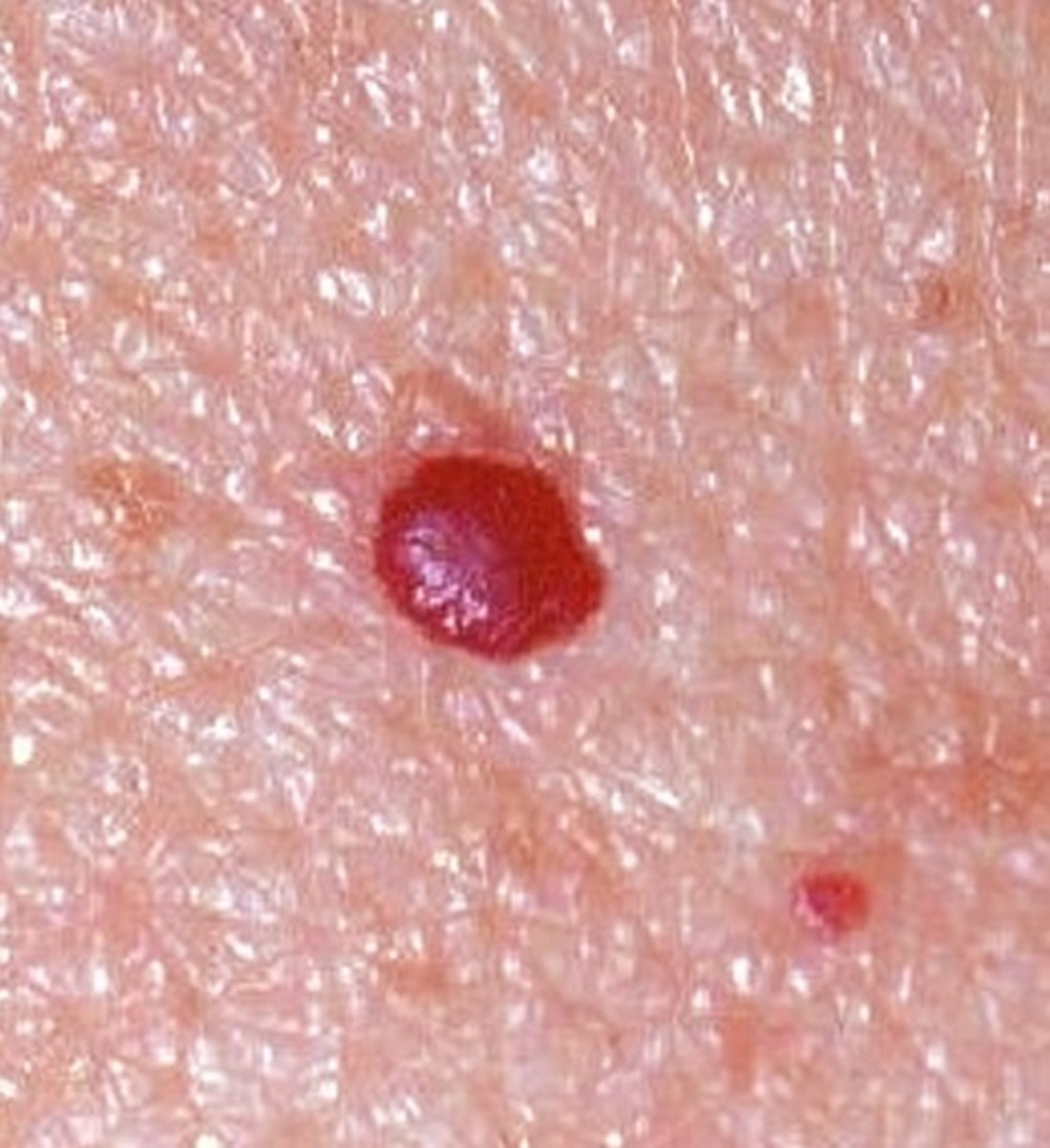
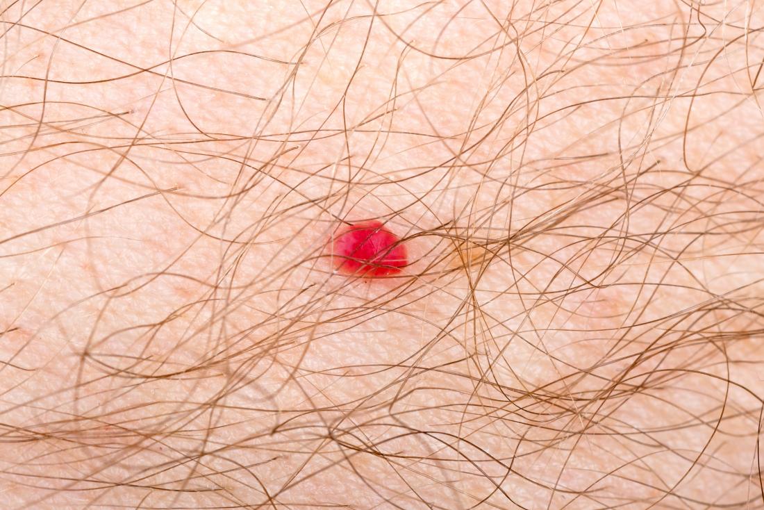
Pictures Symptoms Cherry angiomas get their name from their appearance. Their bright red color occurs due to the dilated capillaries. However, cherry angiomas can be a range of colors and.

Cherry Angiomas Mclean VA & Woodbridge, VA Skin & Laser Dermatology Center
A cherry angioma is a common skin irregularity that causes small, red growths to develop on the skin. The red coloring is caused by numerous, dilated capillaries beneath the skin. The name cherry angioma is derived from the typical cherry-red coloring of these benign skin growths. However, they can actually range in color from red to blue or.

Cherry Angioma Senile Angioma... Academic Dermatology of Nevada
Obencem / Getty Images. Cherry angiomas are extremely common in adults over 30. They are usually found on the torso, though they can appear anywhere, including the arms, legs, chest, and even the scalp. A cherry angioma can be easily removed by a healthcare provider if you desire, but it's not medically necessary. You should never try to remove.
Cherry angioma wikidoc
This is what gives the crusty layer on top of the seborrhoeic keratosis. cherry angioma stock pictures, royalty-free photos & images. Skin problems - Seborrhoeic Keratosis and Cherry Angioma. A senior man's body with a seborrhoeic keratosis, cherry angioma and freckles. A seborrhoeic keratosis is a type of noncancerous skin growth sometimes.
Cherry angioma Symptoms, causes, and treatment
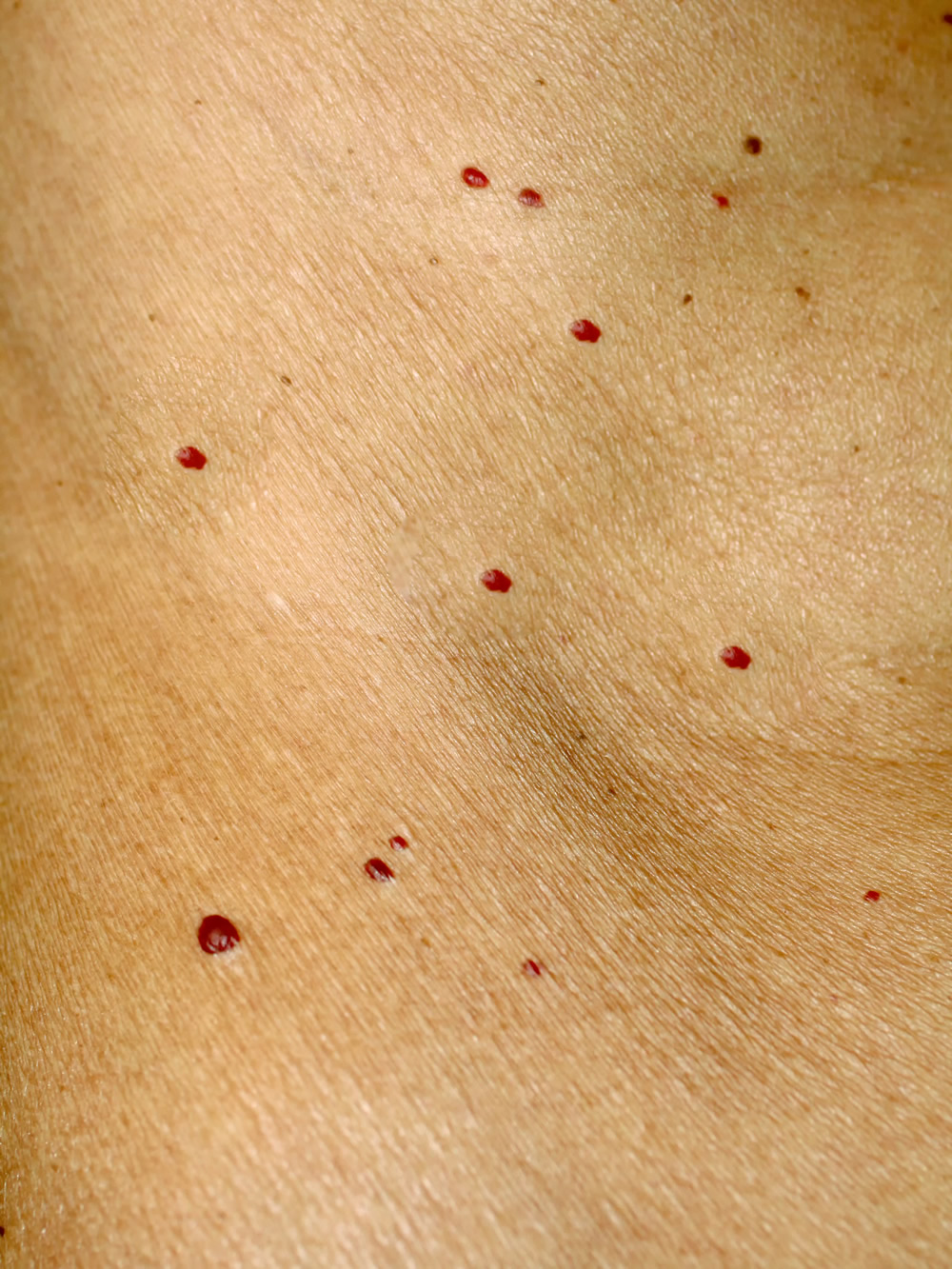
Cherry angiomas are small, pinhead-like lesions on your skin that appear most commonly on your torso, arms and legs of your body. Cherry angiomas are: Round. About 2 millimeters (mm) to 4 mm in size. Light to dark red. The term "cherry" references their color and appearance on the skin, as angiomas typically form in groups. Advertisement
/cherry-angioma-e456f98ada45460db3aeba89281cf3e3.jpg)
Cherry Angioma Symptoms, Causes, Diagnosis, Treatment
Cherry angiomas typically begin as small, flat, bright red spots. They may grow from 1 to 5 mm and become slightly raised. They can be circular or oval in shape. They often grow on the torso, arms.
Cherry Angioma (Cherry Hemangioma) Treatment New York Dr. Michele Green M.D.
Browse Getty Images' premium collection of high-quality, authentic Cherry Angioma stock photos, royalty-free images, and pictures. Cherry Angioma stock photos are available in a variety of sizes and formats to fit your needs.

Drivers Identified in Cherry Angioma Dermatology Advisor
A cherry angioma or cherry hemangioma describes a harmless, benign vascular skin lesion. As seen in the images below, cherry angiomas may occur on any part of the body and removal may be desired for cosmetic purposes.

Cherry angioma (Cherry hemangioma, Senile Angioma, CampbellDe spot) Dermatology Advisor
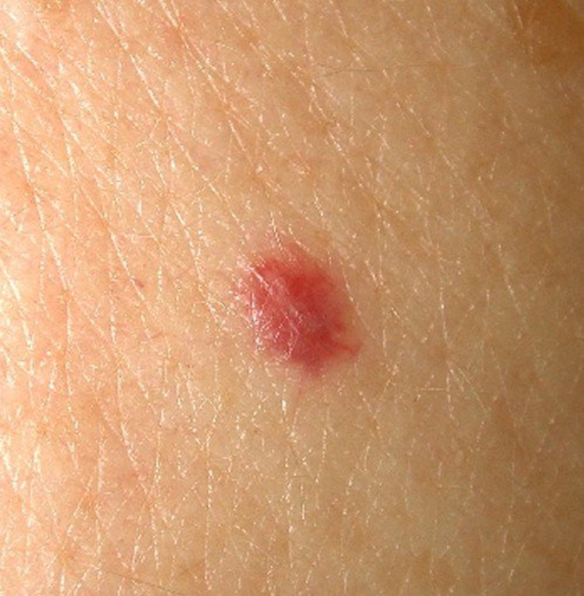
A cherry angioma is a smooth, cherry-red, harmless bump on the skin. They can occur nearly anywhere on the body, and most commonly start appearing around age 40. Cherry angioma quiz Take a quiz to find out if you have cherry angioma. Take cherry angioma quiz What is cherry angioma?
Cherry angioma causes, symptoms, diagnosis & cherry angioma treatment
Laser surgery: different lasers have been used successfully in treatment of cherry and spider angiomas. - Pulsed dye laser (PDL) is the treatment of choice. A spot size should be selected that matches diameter of the angioma. With spider angiomas, the central feeding vessel as well as the surrounding vessels should be treated.
Cherry Angiomas Pictures, Symptoms, Causes, Treatment, Removal HealDove
Cherry angioma images. Authoritative facts about the skin from DermNet New Zealand. DermNet provides Google Translate, a free machine translation service.. Cherry angioma macro and dermoscopic image pairs. Cherry angioma 1 macro. Cherry angioma 1 dermoscopic. Cherry angioma 2 macro. Cherry angioma 2 dermoscopic.

What is Cherry Angioma? Dr. Maksym BreslavetsDr. Maksym Breslavets
Getty Images / AdobeStock Cherry Angiomas Symptoms Cherry angiomas are benign, or noncancerous, skin growths. You may identify a cherry angioma based on characteristics like: Color:.